Feature Article, September 2002
Warning!!!
This article is not for the faint-of-heart as it is very graphic!
If you are the least bit uneasy seeing blood, bodies, or internal body parts do not read this
article. (see full warning)
This month's feature article comes from Brandon Bertolli, who is researching gunshot wounds
in South Africa. Most of this research will be conducted in the adult trauma unit of the Johannesburg Hospital,
Johannesburg, South Africa. This is Mr. Bertolli's third submission and I would like to thank him for taking the time to
submit these interesting case studies to firearmID.com!
Adult Trauma Unit-Case Studies
Johannesburg, South
Africa
The following gunshot cases were encountered by Brandon Bertolli at the
Johannesburg Hospital Trauma unit in 2002.
Case 1
A 14 month old baby sustained a gunshot wound to the right shoulder and head. A
single bullet perforated the shoulder and went on to penetrate the head. The baby was transferred from a small clinic to the
Johannesburg Hospital. The circumstances of the shooting were unknown as no parent accompanied the baby. The baby was intubated.
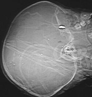
Three wounds were evident: two on the shoulder and one in the right occipital
area of the head. The trajectory of the bullet, based on review of the wounds and the radiological evidence, is only valid if the
baby's neck was flexed at the time of the shooting. The position of the wounds can be seen in this photograph taken after the
shoulder wounds were dressed and the head wound was sutured.
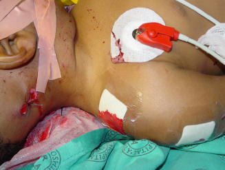
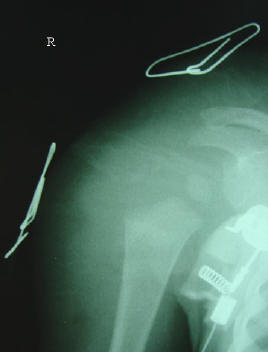
Please note that it is not standard procedure to suture a bullet wound at our
hospital (they are usually dressed like the shoulder wounds), but in this case the wound was bleeding excessively and had to be
sutured. The shoulder X-ray proved that there was no fracture and the skin breaches were marked with paperclips as usual.
The baby then went for immediate CT scan of the brain.
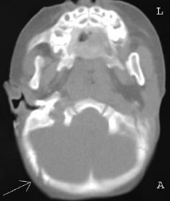
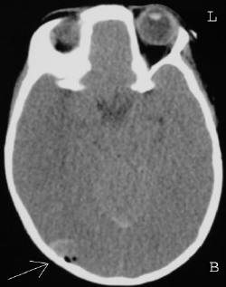
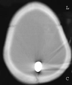
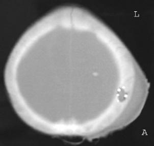
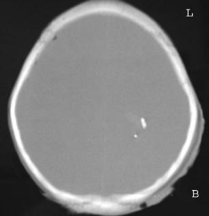
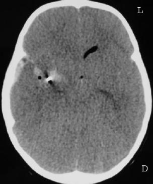
The CT findings were: sub-arachnoid hemorrhage, brain contusion, pneumocephalus
(abnormal pocket of air in the brain signaling a breach of the skull vault, seen on slice B) and a fracture of the occipital bone
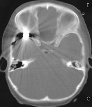
on the right, bordering on the base of the skull (seen on slice A). The bullet track was probably alongside the inner table of the
skull, starting from the right occipital area, leading all the way up to its final resting position at the skull vertex (slice C).
The radiological appearance of the bullet is consistent with that of a full metal jacket bullet. Unfortunately the bleeding and
damage to the brain caused raised intracranial pressure and the baby "coned." Coning is when the pressure inside the
skull due to hematoma or other abnormal collection pressing on the brain, becomes so great that the patient's brain is forced
inferiorly so that the brain stem is trapped in the foramen magnum. The brain stem is vital for controlling all the impulses to
vital organs such as the heart and lungs. If the stem sustains sufficient damage, these impulses are disrupted and the patient
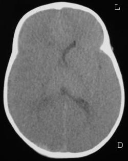
dies. Evidence of raised intracranial pressure can be seen on slice D where the ventricles are compressed on the right. This baby
died within 24 hours of being shot.
Case 2
We were all shocked by the arrival of another baby with a gunshot wound to the
head, just four days after the arrival of the case above. This infant was 20 months old and was being held in his mother's arms at
the time he was shot. It is unclear whether the mother was the target or the infant was the target, because the mother sustained a
minor shoulder wound from a second bullet. She was very vague about the circumstances of the shooting. Even by South African
standards, it is a very grim situation if you have to deal with two pediatric gunshot heads within 4 days of each other.
This baby arrived at the hospital with his mother and was intubated. There was a
wound on the left vertex area of the head, but no exit. Brain matter was seen to ooze from the wound.
There was a struggle to maintain the infant's vital signs and a CT scan of the
brain was performed. Findings: raised intracranial pressure, bullet crossed the midline and was lodged in the right temporal lobe
behind the right orbital wall. Bullet fragments were shed along the bullet track.
The entrance puncture of the skull can be seen in slice A, with fragment
deposition in slice B and the parent fragment rest site in the right temporal lobe, bordering on the right orbit in slice C. There
is evidence of intracranial hemorrhage and raised intracranial pressure on slice D. There is also a bullet fragment evident on
slice D. Unfortunately this infant also died within 24 hours.
Case 3
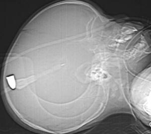
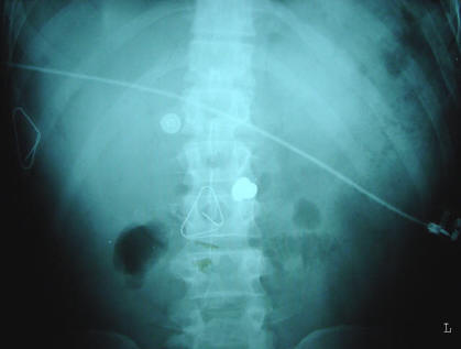
A middle-aged man was hijacked and sustained multiple gunshot wounds to the
abdomen after being pulled out of his vehicle. He was brought to hospital by helicopter, had a single X-ray in the resuscitation
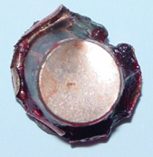
bay and was rushed to theatre for a laparotomy. There were multiple perforating wounds and one penetrating wound with an expanded
intact hollow point bullet lodged in the abdomen. The characteristic shape of the expanded hollow point bullet can be seen on
X-ray. Note that there is another metallic artifact in the area: an ECG electrode.
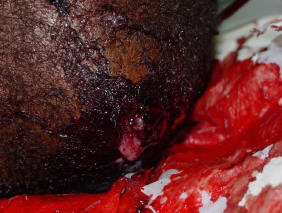
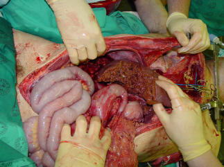
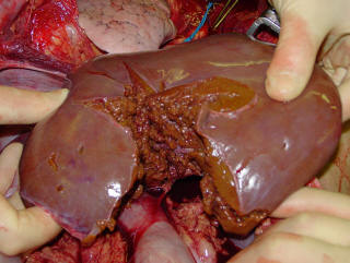
The laparotomy findings were: extensively damaged inferior vena cava, holes in
the stomach, spleen, small and large bowel and a massive cavitating hole in the liver which almost cut the liver in half. The
liver had stellate tears in it from the cavitating rupturing effect of the projectile injury. The I.V.C was so badly damaged that
a shunt had to be put in to return the venous blood from the liver directly to the right atrium of the heart. To this end, a
thoracotomy was performed.
The surgeons battled for two hours to try to save the man, and despite employing
high tech cell-saving machinery and having a spare anesthetist to handle fluids and pressures, the damage was too great and the
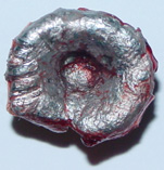
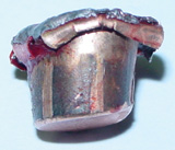
patient did not survive. He had sustained too many injuries at the same time, each one bleeding profusely. The hollow point was
retrieved and the surgeon took care not to handle the bullet with any metal instrument so as not to add tool marks to the surface.
It could be any one of a number of widely available hollow points such as the Winchester JHP. Only detailed analysis will reveal
the type of ammunition and the weapon used to fire it. Preliminary analysis indicates that the bullet is of the 9mm Parabellum
variety. Handgun projectiles can cause cavitating injuries of inelastic organs such as the spleen, kidneys and liver.
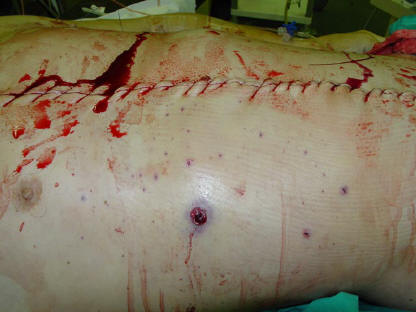
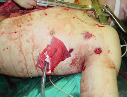
It is obvious from the stipple on the patient's right flank that he was shot at
close range. There were multiple breaches on the left flank and on the back.
The incidence of non-accidental gunshot wounds in Johannesburg has risen sharply
since 1994. The average gunshot victim attendance at the Johannesburg Hospital was 150 per month in 1999. It has now risen to 160
per month (as of statistics available for 2001.)
If anyone has any questions about X-Rays, you can contact me at bbertolli@yahoo.com.
All images seen on this page are copyright © 2002 Brandon Bertolli. All
rights reserved.
|